
by Gertrud U. Rey
The absence of infection with some parasitic worms (also known as helminths) often coincides with the development of asthma, allergy, and autoimmune disorders, suggesting that these worms may have co-evolved a commensal relationship with their human hosts. However, infection with these worms may also lead to an impaired immune response against pathogenic infections by other parasites, bacteria, and some viruses.
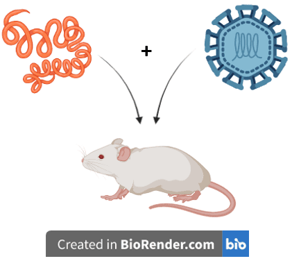
Based on such observations, the authors of a recent study explored the effect of infection with Heligmosomoides polygyrus bakeri (Hpb) – a naturally occurring intestinal roundworm of rodents – on West Nile virus (WNV) infection outcome in mice. WNV is a member of the Flavivirus genus, along with Dengue virus, Zika virus, yellow fever virus, tick-borne encephalitis virus, and Powassan virus. These viruses all infect their human hosts through the bite of an arthropod and ultimately target cells of the central nervous system.
To assess the effect of intestinal Hpb infection on the pathogenesis of WNV, the authors used three groups of mice: the first group was only fed Hpb larvae to initiate an Hpb infection, the second group was only infected with WNV by inoculation in the foot pad, and the third group was infected with both Hpb and WNV. Infection with Hpb alone had no adverse effect on body weight or survival of the mice, and infection with WNV alone caused moderate weight loss and 86% survival. In contrast, infection with both Hpb and WNV led to greater weight loss and only 28% survival. To determine the reason for this enhanced pathogenicity, the mice were euthanized, and different organs were assessed for viral titers. Mice infected only with WNV, or with both WNV and Hpb, had similar levels of virus in the spleen, an organ that filters the blood and produces immune cells in response to the presence of infectious pathogens. However, mice infected with both Hpb and WNV had higher levels of virus in different parts of the colon and the mesenteric lymph nodes, which serve as a firewall to prevent systemic spread of microorganisms. The presence of such high viral titers in these regions suggested that co-infection with Hpb and WNV leads to the infection of cells in gut-proximal places.
To determine which gut cells are targeted by WNV during a Hpb/WNV co-infection, the authors stained pieces of gut tissue from Hpb/WNV co-infected mice with a combination of fluorescent antibodies that bind WNV antigens or neuronal cell markers. Analysis of these tissues by fluorescence microscopy revealed that co-infected animals have higher levels of viral antigens in intestinal neurons compared to control animals infected only with Hpb or WNV, suggesting that infection with both Hpb and WNV increases the frequency of infection of gut neuronal cells. Microscopic analysis also revealed certain gut abnormalities in co-infected animals, including changes in the composition of intestinal villi, which are small, finger-like projections that extend into the lumen of the small intestine. Co-infected animals had shortened villi, a phenomenon typically associated with pathology and inflammation. Co-infected animals also had commensal bacteria inside their spleens, so it is possible that the shortened villi enabled translocation of the bacteria across the epithelial gut barrier, which normally restricts the entry of harmful substances by separating the intestinal organs from the external environment.
The spleens and brains of Hpb/WNV co-infected mice also contained markedly reduced numbers of WNV-specific CD8+ T cells, which usually contribute to the clearance of WNV infection. The authors reasoned that the lack of CD8+ T cells was likely due to bacterial invasion, which may impact the activation of key immune cell types involved in triggering the differentiation of virus-specific CD8+ T cells. Remarkably, treatment of Hpb/WNV co-infected mice with antibiotics reversed these kinetics, leading to increased levels of WNV-specific CD8+ T cells, a result that correlated with diminished bacterial levels in the blood and spleen. This observation suggested that bacterial invasion of lymphoid tissues is largely responsible for impaired WNV-specific CD8+ T cell responses during co-infection with Hpb and WNV.
The clearance of helminth infections is typically mediated by the activation of “type 2” immune responses, which are characterized by the production of various cytokines, including IL-4. Binding of IL-4 to its cognate cell surface receptor activates STAT6, a cytoplasmic transcription factor that ultimately signals the activation of macrophages and differentiation of T cells. To determine if these type 2 immune responses contribute to the lethality observed after co-infection with Hpb and WNV, the authors performed the next set of experiments on “STAT6 null” mice, which lack the STAT6 gene. Interestingly, STAT6 null mice co-infected with Hpb and WNV did not have any of the increased lethality observed in wild type mice. Hpb/WNV-co-infected STAT6 null mice also had reduced overall viral levels and no gut abnormalities characteristic of enhanced pathogenesis, suggesting that STAT6 signaling is needed for development of enhanced disease.
To determine the relevance of IL-4 in the STAT6 signaling cascade, wild type mice were infected with WNV alone, followed by administration of IL-4. This regimen led to increased pathogenicity like that observed in Hpb/WNV co-infected mice, including decreased survival, weight loss, and higher WNV burden in the brain and gut. In contrast, the same WNV/IL-4 regimen given to STAT6 null mice did not produce the same pathogenicity, suggesting that the effects of IL-4 are dependent on the presence of STAT6.
Type 2 immune responses in the gut are initiated by tuft cells, which are specialized cells of the intestinal epithelium that are capable of multiplying in response to the presence of certain parasites. To see whether tuft cells are needed for Hpb-mediated enhanced WNV disease, the authors infected “tuft null” mice (mice lacking tuft cells) with both Hpb and WNV. These mice did not develop any of the effects characteristic of enhanced pathogenesis, suggesting that tuft cells are needed for development of enhanced disease.
Tuft cells selectively express a receptor for succinate, a metabolite needed for energy production. To see whether succinate sensing is involved in enhanced WNV disease, wild type mice were administered succinate and were then infected with WNV. All these mice developed enhanced disease, even in the absence of Hpb infection. However, the same succinate/WNV regimen in tuft null mice, or in mice expressing tuft cells but lacking succinate receptors, did not lead to enhanced disease. These experiments further confirm that succinate receptor-expressing tuft cells are needed to initiate the signaling pathway leading to enhanced disease. Collectively, these results suggest that Hpb-mediated activation of type 2 immune responses in intestinal epithelial cells depends on tuft cells and might be initiated by succinate sensing.
Similar enhanced disease characteristics were also observed after co-infection of mice with Hpb and Zika virus or Powassan virus, suggesting that co-infection with helminths may enhance the disease severity of multiple human flaviviruses. Although these parasitic worm/flavivirus co-infections are currently more common in tropical regions, their prevalence may spread with increasing global climate changes/warming. This study outlines a fascinating aspect of the interface between parasitic and viral infections that warrants further exploration.
[For a more detailed discussion of this paper, I recommend This Week in Parasitism episode 194. The material in this blog post is also covered in this video.]
The absence of infection with some parasitic worms: is curious that causes asthma, allergy, and autoimmune disorders. This is such an interesting post that people need more to see.