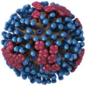

Intranasal inoculation of mice with the PR8 strain of influenza virus leads to injury of both the lung and the intestinal tract, the latter accompanied by mild diarrhea. While influenza virus clearly replicates in the lung of infected mice, no replication was observed in the intestinal tract. Therefore injury of the gut takes place in the absence of viral replication.
Replication of influenza virus in the lung of mice was associated with alteration in the populations of bacteria in the intestine. The numbers of segmented filamentous bacteria (SFB) and Lactobacillus/Lactococcus decreased, while numbers of Enterobacteriaceae increased, including E. coli. Depletion of gut bacteria by antibiotic treatment had no effect on virus-induced lung injury, but protected the intestine from damage. Transferring Enterobacteriaceae from virus-infected mice to uninfected animals lead to intestinal injury, as did inoculating mice intragastrically with E. coli.
To understand why influenza virus infection in the lung can alter the gut microbiota, the authors examined immune cells in the gut. They found that Mice lacking the cytokine IL-17A, which is produced by Th17 helper T cells, did not develop intestinal injury after influenza virus infection. However these animals did develop lung injury.
Th17 cells are a type of helper T cells (others include Th1 and Th2 helper T cells) that are important for microbial defenses at epithelial barriers. They achieve this function in part by producing cytokines, including IL-17A. Th17 cells appear to play a role in intestinal injury caused by influenza virus infection of the lung. The number of Th17 cells in the intestine of mice increased after influenza virus infection, but not in the liver or kidney. In addition, giving mice antibody to IL-17A reduced intestinal injury.
There is a relationship between the intestinal microbiome and Th17 cells. In mice treated with antibiotics, there was no increase in the number of Th17 cells in the intestine following influenza virus infection. When gut bacteria from influenza virus-infected mice were transferred into uninfected animals, IL-17A levels increased. This effect was not observed if recipient animals were treated with antibiotics.
A key question is how influenza virus infection in the lung affects the gut microbiota. The chemokine CCL25, produced by intestinal epithelial cells, attracts lymphocytes from the lung to the gut. Production of CCL25 in the intestine increased in influenza virus infected mice, and treating mice with an antibody to this cytokine reduced intestinal injury and blocked the changes in the gut microbiome.
The helper T lymphocytes that are recruited to the intestine by the CCL25 chemokine produce the chemokine receptor called CCR9. These CCR9 positive Th cells increased in number in the lung and intestine of influenza virus infected mice. When helper T cells from virus infected mice were transferred into uninfected animals, they homed to the lung; after virus infection, they were also found in the intestine.
How do CCR9 positive Th cells from the lung influence the gut microbiota? The culprit appears to be interferon gamma, produced by the lung derived Th cells. In mice lacking interferon gamma, virus infection leads to reduced intestinal injury and normal levels of IL-17A. The lung derived CCR9 positive Th cells are responsible for increased numbers of Th17 cells in the gut through the cytokine IL-15.
These results show that influenza virus infection of the lung leads to production of CCR9 positive Th cells, which migrate to the gut. These cells produce interferon gamma, which alters the gut microbiome. Numbers of Th17 cells in the gut increase, leading to intestinal injury. The altered gut microbiome also stimulates IL-15 production which in turn increases Th17 cell numbers.
It has been proposed that all mucosal surfaces are linked by a common, interconnected mucosal immune system. The results presented in this study are consistent with communication between the lung and gut mucosa. Other examples of a common mucosal immune system include the prevention of asthma in mice by the bacterium Helicobacter pylori in the stomach, and vaginal protection against herpes simplex virus type 2 infection conferred by intransal immunization.
Do these results explain the gastrointestinal symptoms that may accompany influenza in humans? The answer is not clear, because influenza PR8 infection of mice is a highly artificial model of infection. It should be possible to sample human intestinal contents and determine if alterations observed in mice in the gut microbiome, Th17 cells, and interferon gamma production are also observed during influenza infection of the lung.

Pingback: How influenza virus infection might lead to gas...
Pingback: TWiV 316: The enemy of my enemy is not my friend